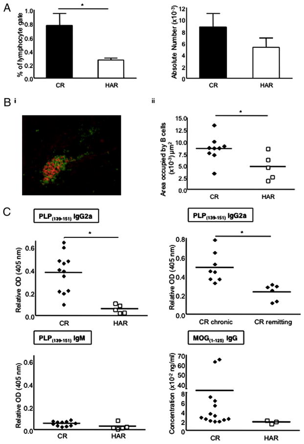
FIGURE 6.
Evaluation of CNS-resident B cells and CNS Ag-specific Ab responses during the postacute phase of EAE. A, Mean frequency and numbers (mean and SE) of B cells in the spinal cords of postacute-phase EAE mice. Results are representative of four to six mice per substrain. B, i, A representative image of a B cell follicle in the brain meninges taken from a CR SJL/J mouse during the postacute phase of EAE (red, B220 stain; green, CD4 stain; ×200 magnification). ii, Quantification of the B cell areas in CNS meningeal follicles from postacute-phase mice (day 21). C, Relative α-PLP139–151 and α-MOG1–125 Ab titers in serum of mice (CR versus HAR or CR chronic versus CR remitting) during the postacute phase of disease. Serum titers from both postacute time points (days 21 and 29) were similar. Five to 15 mice were tested in each group. *p < 0.05.

