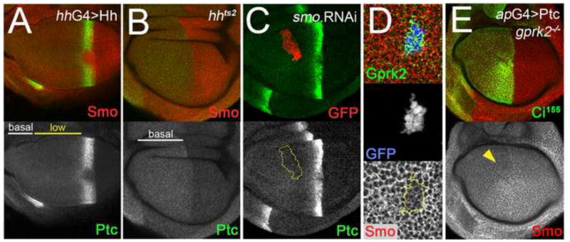
Fig. 7. Ptc protein reflects the Hh ligand gradient, and regulates Smo accumulation independently of Gprk2.
(A) Wing disc in which the range of Hh ligand diffusion was increased by overexpressing Hh in the posterior compartment (driven by hh-Gal4). Ptc is in green, Smo in red. The “low-Ptc” region extends through most of the anterior compartment. (B) hhts2 mutant wing disc in which functional Hh ligand was eliminated after culture for 16 h at the restrictive temperature (29°C). Ptc is in green, Smo in red. The “low-Ptc” region is absent. (C) Wing disc with a clone expressing a dsRNA targeting Smo situated in the “low-Ptc” region. The clone is marked by co-expression of GFP (red channel). The levels of Ptc (green channel) in the clone are unchanged. (D) Closeup view of a posterior compartment clone of ptcS2 homozygous mutant cells co-expressing GFP (blue channel) and Gprk2 (green channel), generated using the MARCM system. Smo levels (red channel) are lower in Gprk2-expressing ptc mutant cells than in surrounding wild-type cells. (E) gprk2KO/gprk2KO mutant wing disc in which Ptc was overexpressed in the dorsal compartment with ap-GAL4. Hh signaling was inhibited by Ptc expression, as evidenced by the loss of Ci155 stabilization in dorsal cells (green channel). Smo levels (red channel) were reduced throughout the dorsal compartment, most notably in anterior compartment cells that would otherwise show ectopic accumulation of the protein (yellow arrowhead).

