
Figure 4. Protein phosphorylation or dephosphorylation after lithium treatment.
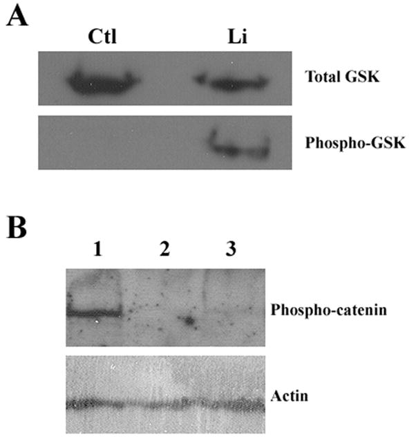
FLS cells were serum-starved for 48 hrs and then were treated with 100 mM lithium chloride for 12 hrs. Cells were collected, and proteins were fractionated by SDS-PAGE. A, Western blot analysis of GSK-3β. Upper panel, total GSK-3β as revealed by using an antibody raised against unphosphorylated protein. Lower panel, phosphorylated GSK-3β as revealed by using an antibody specific for the phopsho-serine (Ser-9) epitope of the protein. Ctl, control cells treated with fresh serum-free medium; Li, cells treated with lithium. B, Western blot analysis of β-catenin. Upper panel, phosphorylated β-catenin detected using an antibody specific for phosphorylated (Thr-41/Ser-37/Ser-33) β-catenin protein. Lower panel, actin proteins detected using anti-β-actin antibody, indicating protein loading. Lane 1, cells treated with fresh serum-free medium; Lane 2, cells treated with lithium; Lane 3, cells treated with GSK-3β inhibitor VII.