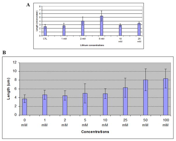
Figure 8. Dose response for lithium in two neuronal cell types.
Cells were serum-starved for 48 hrs and then were treated with lithium at the concentrations indicated in the figure. A, PC-12 cells; B, astrocytes. Shown are average lengths of 150 primary cilia obtained from three independent experiments, with error bars indicating standard deviation.

