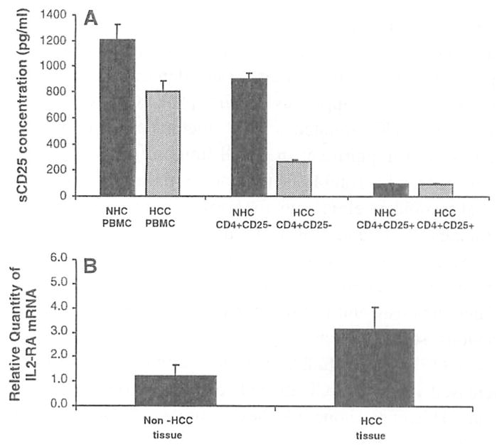
Fig. 5.

Sources of sCD25. a PBMC, CD4+CD25− T cells, and CD4+CD25+ T cells from NHC (n = 5) and patients with advanced HCC (n = 5) were stimulated with PHA and analyzed for their ability to secrete sCD25 into culture which was quantified by ELISA to characterize the cellular source of sCD25. PBMC (1.205 ± 117 pg/ml) and the CD4+CD25− T cell cultures (898 ± 44 pg/ml) from NHCs produced higher levels of sCD25 during the culture compared to CD4+CD25+ T cells (131 pg/ml, lower limit of detection of ELISA). In contrast, in the HCC cultures, the CD4+CD25− T cell cultures produced low levels of sCD25 (259 ± 19 pg/ml) while the PBMC cultures secreted higher levels of sCD25 (803 ± 84 pg/ml). This analysis suggested that the major source of sCD25 in the NHC cultures was the CD4+CD25− T cell fraction while in the HCC cultures non-CD4 T cells and/or antigen presenting cells may be the predominant source, b Real-time PCR was performed after isolating IL2-RA mRNA from liver tissue with HCC and without to determine if tumor is a source of sCD25. The mean level of IL2-RA mRNA from liver tissue with HCC was compared to liver tissue without HCC from the same patient (n = 6). There was a threefold increase in the IL2-RA mRNA found in the liver tissues with tumor when compared to sites without tumor (P < 0.05). Messenger RNA expression for IL2-RA was standardized to GAPDH mRNA expression and expressed as the fold-difference compared to normal for each tissue sample
