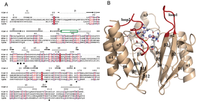
Figure 1. Overall structure of NDM-1 and its sequence alignment with its homologue proteins.
A. Sequence alignment of NDM-1 with VIM-4, FEZ-1 and CphA. The second structure assignment of VIM-4 is labeled on the top of the sequences. Black quadrangles indicate residues that coordinate with Zn2+(I), while black circles indicate residues that coordinate with Zn2+(II). The residues composing of the loop1 of NDM-1 is labeled in green box. B. Cartoon representation of the overall structure of NDM-1 is in light orange color. The loop1, loop2 and the insertion are colored in red, the conserve residues coordinating with zinc ions are represented as sticks in gray color, and the zinc ions shown as spheres.

