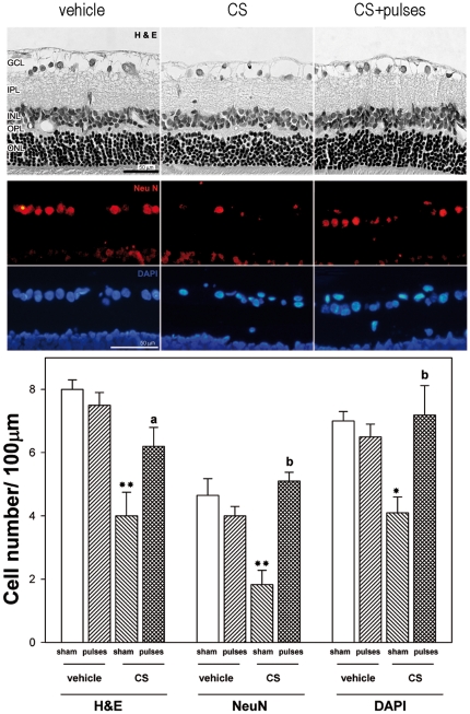
Figure 3. Retinal histology examination after 10 weeks of ocular hypertension.
Upper panel: Representative photomicrographs of retinal sections stained with hematoxylin and eosin from a vehicle-injected eye, and a hypertensive eye without or with pulses of ischemia. Note the diminution of GCL cells in the eye injected with CS without ischemia pulses. The application of ischemia pulses preserved this parameter. The other retinal layers showed a normal appearance in all groups. Middle panel: Immunohistochemical detection of NeuN-positive neurons in the GCL from a vehicle-injected eye, a hypertensive eye without or with ischemia pulses. A strong NeuN-immunostaining (red) was confined to ganglion cells in the GCL. The number of NeuN positive ganglion cells was lower in hypertensive eyes without ischemia pulses than in vehicle- injected eyes, whereas the application of ischemia pulses in CS-injected eyes increased NeuN-immunostaining. A similar profile was observed for cell nuclei counterstained with DAPI (blue). Lower Panel: cell count in the GCL evaluated by H&E staining, NeuN immunostaining, and DAPI labeling. By all these methods, a significant decrease of the number of cells in the GCL was observed in CS- injected eyes without ischemia pulses as compared with vehicle-injected eyes (sham), whereas ischemia pulses significantly preserved this parameter in CS-injected eyes. Scale bar: Upper panel = 50 µm; Middle panel = 50 µm. Data are the mean ± SEM (n = 5 animals per group). * p<0.05, ** p<0.01 vehicle injected eyes without ischemia pulses (sham), a: p<0.05, b: p<0.01 versus CS-injected eyes without ischemia pulses (sham), by Tukey's test. GCL, ganglion cell layer; IPL, inner plexiform layer; INL, inner nuclear layer; OPL, outer plexiform layer; ONL, outer nuclear layer.

