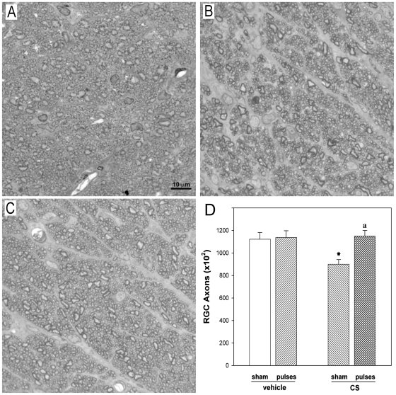
Figure 5. ONH sections from a control or a CS-treated eye without or with ischemia pulses.
(A) Healthy, intact control optic nerve. Note the homogeneity of the staining. In vehicle-injected eyes, individual axons were generally uniform in shape, rounded and packed together tightly to form the fibers of the healthy nerve. In CS-treated eye without ischemia pulses (B) a less stained area indicates a nerve alteration. Disease in individual axons was characterized by axonal distention and distortion that resulted in a departure from the circular morphology of normal axons. In contrast, a conserved structure of the ONH was observed in the CS-treated eye with ischemia pulses (C). Toluidine blue. (D) Number of axons in eyes injected with vehicle or CS without or with ischemia pulses. A significant decrease in the axon number was observed in CS- injected eyes without ischemia pulses as compared with vehicle-injected eyes (sham), whereas ischemia pulses significantly preserved this parameter. Scale bar: 10 µm. Data are mean ± SEM (n = 5 eyes/group) *p<0.05 vehicle injected eyes without ischemia pulses (sham), a: p<0.05 versus CS-injected eyes without ischemia pulses (sham), by Tukey's test.

