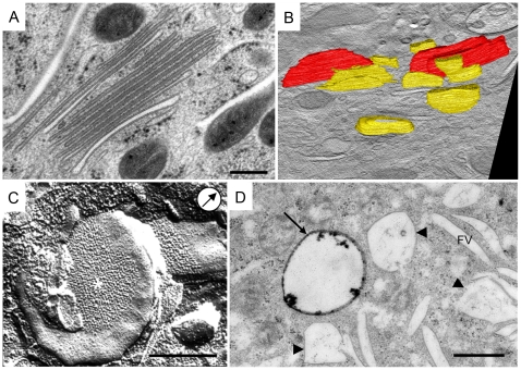
Figure 4. mFVs present the final stage of urothelial plaque formation.
A) On the micrograph are shown mFVs organized into a stack. mFVs have a narrow intravesicular lumen and un-dilated rims. B) Projection of 3-D model shows difference in size and shape between bigger iFV (yellow) and mFVs (red). C) mFVs have urothelial plaques (*) positioned centrally. Note flattened, coin-like shape of the mFV. Arrow in circle indicates direction of Pt shadowing. D) After two hours of endocytosis, the HRP reaction product is present in a multivesicular body (arrow), while the majority of multivesicular bodies (arrow-heads) and all of the FVs remain un-labelled. Bars 500 nm.

