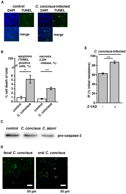
Figure 4. Effects of C. concisus on epithelial apoptosis.
( A ) Apoptotic effects of C. concisus on confluent HT-29/B6 cells visualized with TUNEL staining and fluorescence microscopy 48 h after infection, the number of cell rosette formations was increased, condensed or fragmentized nuclei were visible, and a more generalized loss of cells was observed. ( B ) After 48 h of incubation with C.concisus TUNEL staining of confluent HT-29/B6 monolayers revealed an increase in the number of apoptotic cells and LDH release was measured as a sign of necrosis induction (Student's t-test with **P<0.01; ***P<0.001; each n = 5). ( C ) Increased apoptosis was indicated by caspase-3 activation 48 h after infection. Band intensity of pro-caspase-3 (35 kDa) was significantly diminished (P<0.05, n = 7). C. jejuni ATCC 33560 served as positive control. ( D ) After 48 h of incubation with C. concisus from oral or fecal source apoptotic cell number was equal in TUNEL staining of confluent HT-29/B6 monolayers (n.s., n = 5). ( E ) For verification of C. concisus-induced apoptosis effects on barrier function, HT-29/B6 monoalyers were pre-treated with 50 µM Z-VAD (N-benzyloxycarbonyl-Val-Ala-Asp-fluoromethyl-ketone), a caspase inhibitor that completely blocks caspase-mediated cell death. Transepithelial resistance was partially although not completely reconstituted by Z-VAD. Isolates were grouped depending on source in “oral” and “fecal” as listed in table 1.

