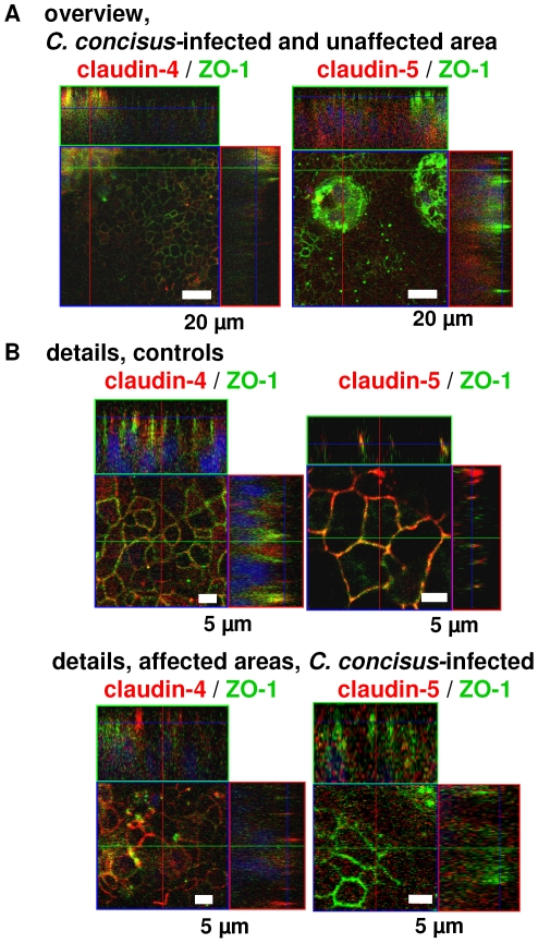
Figure 6. Apoptosis and tight junctions.
Confocal laser-scanning microscopy of HT-29/B6 monolayers revealed patchy distributed areas with tight junction changes and apoptotic events as well as largely unaffected areas in ( A ) overviews with low magnification and ( B ) details with higher magnifcation. Immunostaining with green signal for ZO-1; and claudin-4 and -5 marked red; merge appears yellow. Nuclei are stained blue with DAPI.

