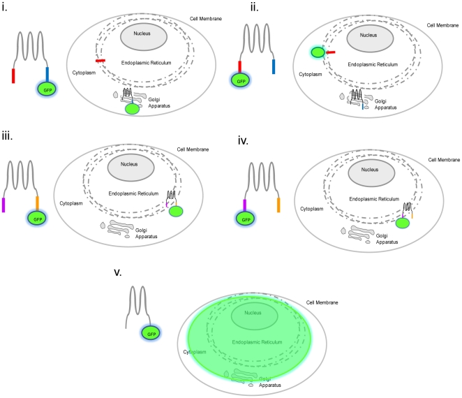
Figure 7. Schematic diagram of a model for localization of ZnT5.
Variant A is expressed as a pro-protein that undergoes cleavage in the ER to remove an N-terminal signal peptide, which is retained in the ER, while the mature, processed protein trafficks to the Golgi apparatus. In contrast, variant B is retained in the ER. Thus, when variant A is detected by virtue of a GFP (or epitope) tag attached to the C-terminus, the signal is observed at the Golgi apparatus (i). If variant A is detected by virtue of a GFP (or epitope) tag attached to the N-terminus, however, the signal is observed at the ER (ii). In the case of variant B, which does not undergo any proteolytic cleavage, the signal is observed at the ER irrespective of whether the GFP (or epitope) tag is at the C-terminus (iii) or N-terminus (iv). Retention within the ER/Golgi apparatus requires the region 525 to 666, so a diffuse signal is observed when the protein is detected by virtue of a GFP (or epitope) tag attached (at either the N- or C-terminus) to a truncated protein lacking this region (v). Note that there is no intention that the toplogical representation of the protein is accurate.

