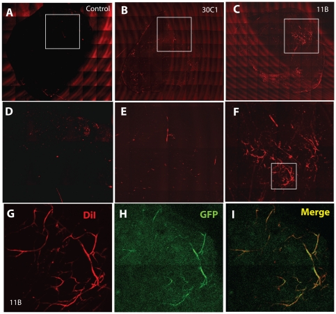
Figure 7. Clonal heterogeneity of CPC vasculogenic properties in vivo.
A–C. Vascular differentiation of SDF-1-producing CPCs in vivo. Whole mount scan of Matrigel plugs containing no cells (control, A), low SDF-1 clone 30C1 (B) or high SDF-1 clone 11B (C). D–F: Enlargements of boxed areas as shown. G, H: Vascular structures in CPC-seeded Matrigel plugs. Representative fluorescence microscope images of plugs containing clones 30C1 (G) or 11B (H) under identical in vivo conditions. I. Overlap of DiI-stained and GFP-expressing blood vessels. Additional images are provided in Figure S4.

