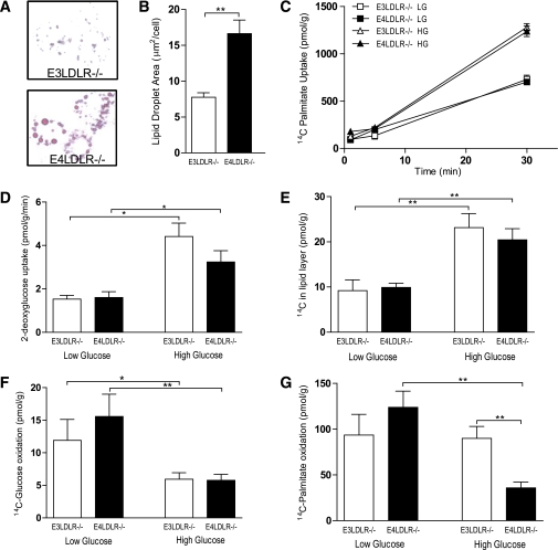
FIG. 5.
Metabolic analyses of primary hepatocytes. A: Primary hepatocytes from E3LDLR−/− or E4LDLR−/− mice were cultured for 72 h in high glucose (25 mmol/L) media and stained with Oil Red O to highlight lipid droplets. B: Total lipid droplet area per cell was quantified by measuring 50 randomly chosen cells from four separate cultures per group. C: Fatty acid uptake was estimated by counting intracellular radiation after incubating hepatocytes for 1 min with 2 µCi/mL [14C]palmitate. D: Glucose uptake was measured after a 10-min incubation with 1 µCi/mL 3H-2-deoxyglucose after starving cells for 2 h. E: DNL was measured by counting radiation in the lipid layer after 24-h incubation with [14C]glucose. F and G: Oxidation of [14C]glucose (F) and [14C]palmitate (G) was measured by trapping 14CO2 during a 3-h incubation using a customized, self-contained CO2 trap (n = 4–12 wells per trial, three trials per group). *P < 0.05 and **P < 0.01. (A high-quality color representation of this figure is available in the online issue.)

