FIG. 6.
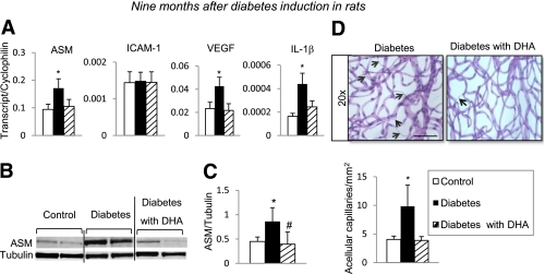
Effect of DHA-supplemented diet on long-term diabetes-induced degenerative changes in rat retina. Retinas isolated 9 months after induction of diabetes were analyzed by quantitative PCR and immunobloting for inflammatory/angiogenic molecule expression. Quantitative PCR analysis of ASM, ICAM-1, VEGF, and IL-1β (A) of retinas isolated from rats subjected to a standard diet (control, white bar; diabetic, black bar) or a DHA-enriched diet (diabetic, striped bar) is shown. The results are means ± SE from one set of animals, with five to eight animals in each group. *P < 0.05 compared with control. Immunoblot (B) and quantitative analysis (C) of ASM protein levels in retinas isolated from control rats on standard diet and diabetic rats on standard and DHA-enriched diet. The results are means ± SD of four animals in each group, performed in triplicate. *P < 0.05 compared with control; #P < 0.05 compared with diabetes. D: Retinal vasculature from control, diabetic, or diabetic supplemented with DHA animals was prepared using trypsin digestion and stained with hematoxylin and periodic acid–Schiff. Dramatically increased number of acellular capillaries (black arrows) was observed in retinal vasculature isolated from diabetic animals; however, diabetic animals supplemented with DHA were protected from acellular capillaries formation. D: Quantification of the total number of acellular capillaries. The results are means ± SD from one set of animals, with eight animals per group. At least eight fields of retina were counted in duplicates by two independent investigators. *P < 0.05 compared with control; black bar = 110 μm. (A high-quality digital representation of this figure is available in the online issue.)

