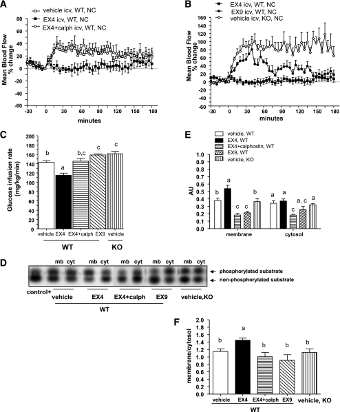
FIG. 1.
Brain GLP-1 signaling recruits PKCs to control femoral artery blood flow and whole-body GIR in WT mice during hyperinsulinemic-hyperglycemic clamp studies. A and B: Mean arterial blood flow in percentage changes over baseline during a hyperinsulinemic-hyperglycemic clamp where vehicle, Ex4, Ex9, or Ex + calphostin (calph) were infused into the brain of WT (A and B) or Glp1r−/− (knock-out [KO]) (B) mice. C: Whole-body GIRs simultaneously recorded in the same mice. Data are means ± SE, n = 5–8 mice per group. D: Gels showing the phosphorylated and nonphosphorylated PKC substrates in the membrane (mb) and cytosolic (cyt) fractions isolated from the hypothalamus of WT or Glp1r−/− mice clamped under similar conditions. E and F: Quantification of PKC activity in AUs and in the membrane-to-cytosol ratio. Differences between groups for the activities assessed in the membrane or cytosolic fractions were analyzed separately. Data with different superscript letters are significantly different (P < 0.05) according to the two-way ANOVA test. Differences between GIR were analyzed according to the one-way ANOVA test. icv, intracerebroventricularly.

