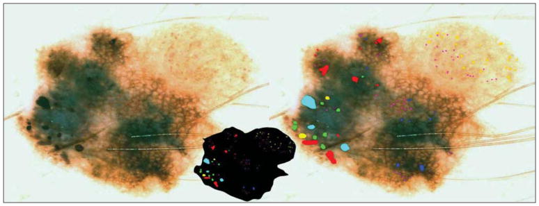
Figure 2.
Malignant melanoma with globules of different sizes and shapes. The globule distribution type is eccentric and shows asymmetric clusters but no peripheral rim. Note that globules marked with green are not necessarily perfectly round or oval but are generally convex. The lesion outline used in determining distribution type is shown in the inset at center. Green indicates classical; red, irregular; yellow, light; lavender, small dot-globule variant; light blue, large; and blue, connect-globule variant.

