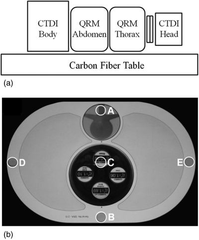
Figure 2.
(a) Measurement setup for thoracic and lumbar spine radiation dose and image quality assessment. The long stack of phantoms approximates a realistic setup that includes dose from x-ray scatter. (b) Photograph of the thoracic phantom. The labels A–E show the location of holes for Farmer chamber placement. For image quality (CNR) measurements, the uniform dosimetry inserts at locations A and C were replaced with the simulated vertebra and set of tissue-simulating inserts as shown in (b). The tissue-equivalent inserts were placed within an acrylic cylinder (clockwise from bottom): cortical bone, B-200 bone, breast, and liver. Since spine surgery is most commonly performed with the patient in a prone position, the phantom was arranged with the spine at the top surface.

