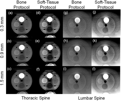
Figure 7.
Tissue-equivalent inserts in the thoracic and lumbar spine phantoms for “Bone Protocol” and “Soft-Tissue Protocol” at various slice thickness. (a–c) Bone detail in the thoracic spine suggests excellent visibility at thin slices with minimal degradation due to quantum noise. (d–f) Soft-tissue scans in the thoracic spine. Reduction in slice thickness shows substantially increased noise. (g–i) “Bone Protocols” in the lumbar spine, showing good bone visibility at thin slices. (j–l) “Soft-Tissue Protocol” scans in the lumbar spine. Increasing slice thickness yields improved soft-tissue visibility through decreasing noise.

