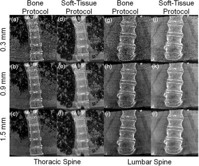
Figure 8.
Thoracic and lumbar spine images of a cadaver scanned using “Bone Protocol” and “Soft-Tissue Protocol” settings, displayed with increasing slice thickness. (a–c) Bone detail visibility for thoracic “Bone Protocol”. (d–f) Soft-tissue thoracic scan protocol. Lung bronchi of 2nd and 3rd generations are easily discernable. (g–i) Low dose “Bone Protocol” acquisition in the lumbar spine. Bone windowing and increased slice thickness (0.9 mm) show good bone detail visualization. (j–l) Soft-tissue visibilities in the lumbar spine for the “Soft-Tissue Protocol (HiRes)” scan protocol. The lumbar paraspinalmuscles and adipose tissue running along the spine, as well as intervertebral discs, can be easily discerned at a slice thickness of 0.9 mm.

