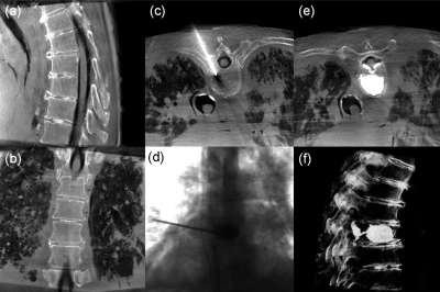
Figure 9.
Example images illustrating CBCT and fluoroscopy in the course of vertebroplasty. (a and b) Sagittal and coronal slices of a CBCT acquired prior to first incision (“Bone Protocol”). (c) Axial CBCT image showing the canula through the right mid-thoracic pedicle (“Bone Protocol”). (d) Single-frame fluoroscopy acquired during cement injection. (e) Axial CBCT image after cement injection (“Bone Protocol”). The cement is easily discernable, as is extravasation and breach of the spinal canal (imparted intentionally to test visualization of such complication). (f) Volumetric display of a CBCT scan acquired following the procedure [“Soft-Tissue Protocol (HiRes)”] for verification of the surgical product. The intentional cement leak into the spinal canal was easily seen as posterior extension of the cement through the basiverterbal vein and into the spinal canal (e and f).

