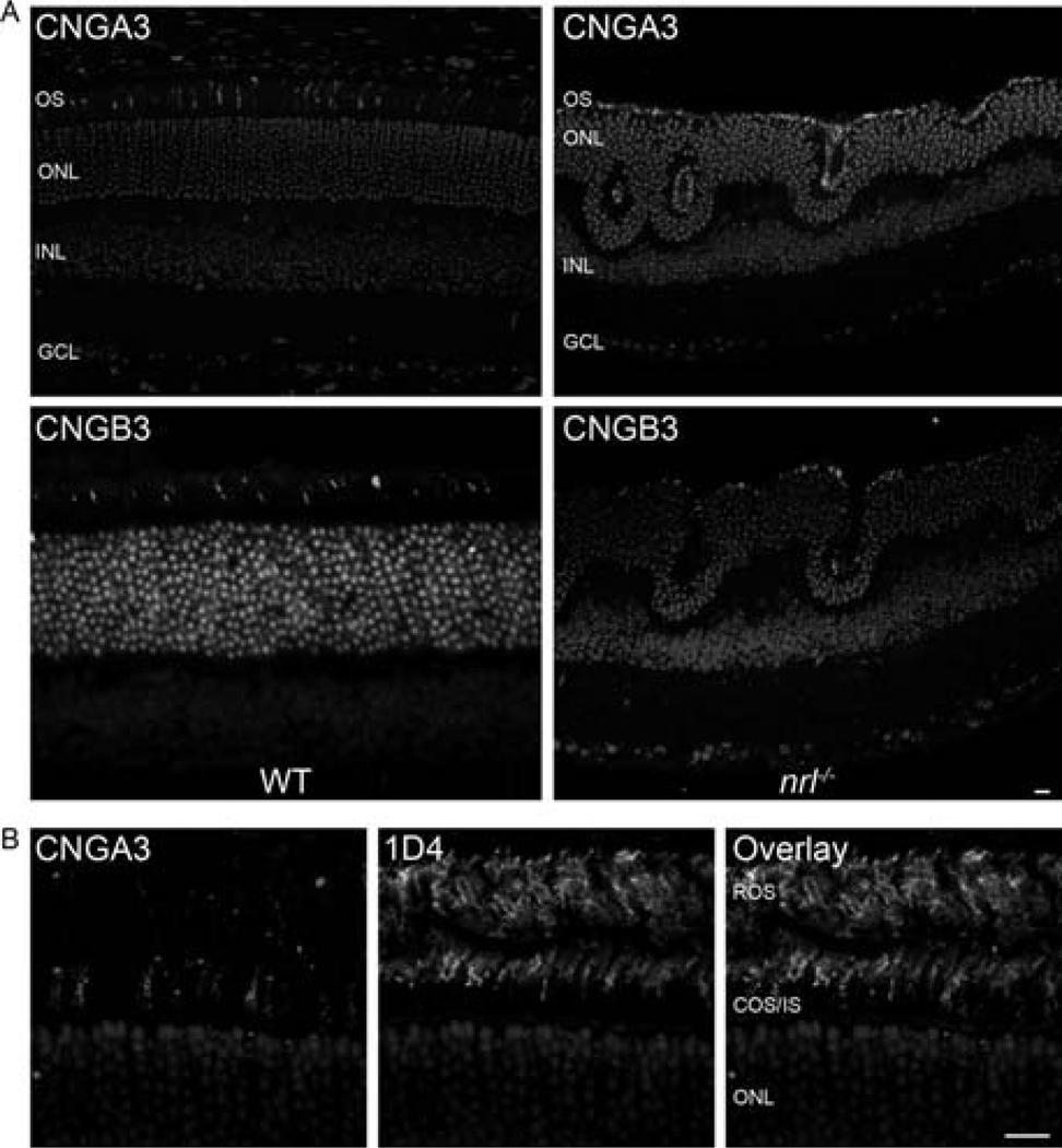
Fig. 8.1.
CNGA3 and CNGB3 is expressed in cones OSs of WT and nrl−/− Retinas; (a) Paraffin embedded sections from P30 WT or nrl−/− mice were stained with CNGA3 and CNGB3 polyclonal antibodies and visualized using anti-rabbit Cy3 secondary antibody. Sections were counterstained with DAPI to label nuclei. Expression of cone channel subunits is limited to the OS layer in both WT and nrl−/− retina. Scale bar 20 μm. (b) P30 WT paraffin embedded sections were stained with CNGA3 (left) and mAB 1D4 (against rhodopsin-middle). The two proteins do not co-localize (right) indicating that cone CNG is not expressed in rods. Scale bar 10 μm. OS, outer segment; ONL, outer nuclear layer; INL, inner nuclear layer; GCL, ganglion cell layer

