Fig. 3.
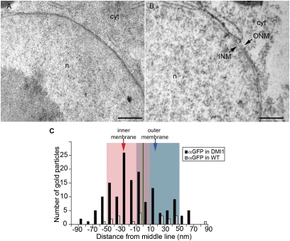
M. truncatula DMI1 preferentially localizes to the inner nuclear membrane. (A) Immunogold labeling of nontransgenic M. truncatula root samples using anti-GFP antibody; few nonspecific signals were detected. (B) Immunogold labeling of DMI1::GFP-expressing transgenic hairy root cells using anti-GFP antibody; signals were detected on both inner and outer nuclear membranes. (C) Quantitative analysis of the relative abundance of DMI1 in the nuclear membranes. Arrows in B indicate the inner (INM) and outer (ONM) nuclear membranes. Black bars are DMI1-GFP transformed roots, grey bars are control roots. n, nucleus; cyt, cytoplasm. (Scale bar, 500 nm.)

