Fig. 5.
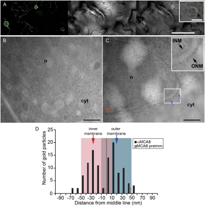
Localization of MCA8 in root epidermal cells of M. truncatula. (A) Transient expression of a truncated version of MCA8 containing the predicted NLS and three transmembrane domains. The GFP signal is concentrated in the nuclear envelope, and some signal can be seen in the ER. Left: Fluorescent image; Center: light microscopic image; Right: merge. Inset: Detail of a nucleus with GFP signal restricted to the nuclear envelope. (B) Immunogold labeling of M. truncatula root cells using preimmune serum for MCA8; no significant signal was observed. (C) Immunogold labeling of M. truncatula root cells using an MCA8 antibody; signals were detected on the nuclear membranes. Inset: Nuclear envelope showing gold particles on the inner (INM) and outer (ONM) nuclear membranes. (D) Quantitative analysis of relative abundance of MCA8 in respective nuclear membranes. MCA8 is equally distributed over both inner and outer nuclear membranes in root epidermal cells of M. truncatula. Red arrow indicates gold particles on the inner nuclear membrane; blue arrow indicates gold particles on the outer nuclear membrane. n, nucleus; cyt, cytoplasm. (Scale bars, 100 μm in A, 15 μm in A Inset, 1 μm in B, 1 μm in C.)

