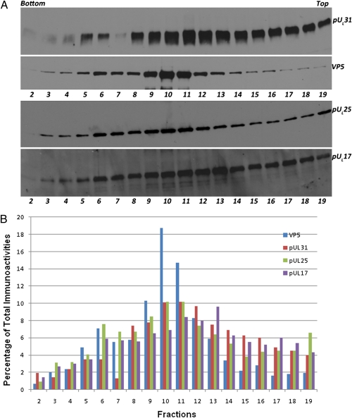
Fig. 2.
Immunoblot of sucrose gradient-fractionated wild-type capsids probed with anti-pUL31, anti-pUL25, or anti-VP5 or anti-pUL17 antibodies. CV1 cells were infected with HSV-1(F) at a multiplicity of infection of 5 pfu/cell. At 20 h after infection cells were collected and lysed. Capsids were pelleted by centrifugation through a sucrose cushion and were then resuspended and separated on a continuous sucrose gradient (Materials and Methods). Approximately 0.5-mL fractions as determined by eye were collected from the bottom of the gradient (fraction 1) to the top (fraction 20) using a Buchler Auto Bensi-Flow IIC gradient collector. Proteins in fractions were TCA precipitated, and pellets were denatured and solubilized in SDS. Fractions 2 through 19 were separated on an SDS polyacrylamide gel and analyzed by immunoblotting, followed by reaction with appropriate conjugates, application of chemiluminescence substrate, exposure to X-ray film, and digital scanning. (A) Top and Upper Middle: Images of the same blot first probed with pUL31 then stripped and reprobed with VP5 antibodies. Lower Middle and Bottom: A second blot containing identical samples probed with pUL25 antibody. Immunoreactivity was then stripped, and the blot was probed with pUL17 antibody. (B) Percentage of immunoreactivity of a given antibody in different fractions as quantified by Image J software.

