Fig. 5.
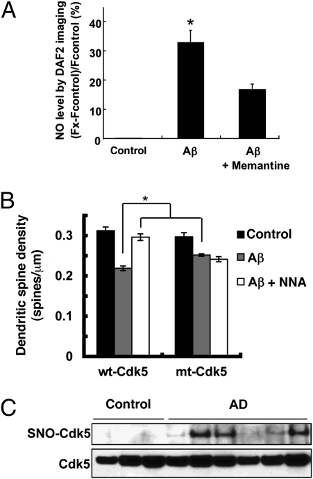
S-Nitrosylation of Cdk5 in response to Aβ in vitro and in brains with Alzheimer’s disease in vivo. (A) Aβ oligomers induce NO in cortical neurons, at least in part, via NMDAR activation. Cortical neurons were preloaded with 4 μM DAF-2 to detect NO and exposed to 5 μM oligomerized Aβ1–42 in the presence or absence of the NMDAR inhibitor memantine (10 μM); similar results were obtained with Aβ25–35, but not with Aβ35–25 or nonoligomerized Aβ1–42. By dynamic light scattering analysis, the oligomerized preparation of Aβ1–42 contained 250 nM of oliogmers. DAF-2 fluorescence was measured using deconvolution microscopy. (B) SNO-Cdk5 mediates in part Aβ-induced dendritic spine loss. Cortical neurons were transfected with pEGFP plus pcDNA3, pEGFP plus wt-Cdk5/p35 or pEGFP plus mt-Cdk5/p35 and then exposed to Aβ25–35 (10 μM for 4 d) or control Aβ35–25. Cells were then fixed and stained with anti-MAP2 antibody to identify neurons. Dendritic spines were visualized by the presence of EGFP protuberances and quantified per micrometer of dendritic length. For A and B, values are mean + SEM (t test with Bonferroni correction, n > 3; *P < 0.05). (C) Postmortem human brain tissues from control subjects or patients with Alzheimer’s disease were subjected to the biotin-switch assay to detect SNO-Cdk5.

