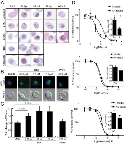
Fig. 4.
Inhibition of PfA-M1 kills parasites via disruption of Hb digestion whereas inhibition of Pf-LAP kills via a distinct mechanism. (A) Parasites were treated with BTA (10 μM), E-64d (10 μM), PNAP (3 μM), and artesunate (10 nM) at concentrations roughly equivalent to their IC99, and followed by Giemsa staining and light microscopy throughout the lifecycle. Scale bar, 5 μm. (B) DV swelling was confirmed by the dose-dependent enlargement of the average DV area. Parasites expressing YFP-tagged plasmepsin II (PMII-YFP), which localizes to the DV, were treated with increasing concentrations of BTA (and 250 nM PNAP) and imaged by fluorescence microscopy. (C) DV swelling was determined to be saturable and quantified by treating the PMII-YFP parasites with increasing concentrations of BTA (PNAP was also used at 250 nM) at midring stage and measuring fluorescent DV area 20 hrs later using a minimum of 10 parasites (+/- standard deviation). (D) Treatment of parasites with BTA in media lacking exogenous amino acids, except for isoleucine, results in a more than twofold decrease in the IC50 of BTA, while parasites treated with PNAP or artesunate show a nonsignificant difference. Shown are representative IC50 plots for each compound in both I-Media (lacking exogenous amino acids except for isoleucine), and AA-Media (containing all natural amino acids). The inlay bar graphs show differences of the mean IC50 of three experiments carried out in triplicate (*P < 0.05, Student’s t test).

