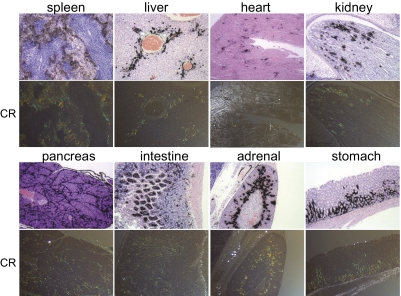
Fig. 5.
Microautoradiography and Congo red staining of amyloid-laden tissues. Mice with advanced AA were injected i.v. with 125I-p5 peptide and euthanized at 1 h pi. Tissues were fixed, and slides were prepared for Congo red (CR) staining or microautoradiography. For microautoradiology, tissue sections were exposed to photographic emulsion for 96 h before being developed and counterstained with H&E. Congo red staining of amyloid appears as green/yellow birefringent material in polarized light microscopy. (Magnification: 80×.) For display, the images were scaled to 10% of original size.

