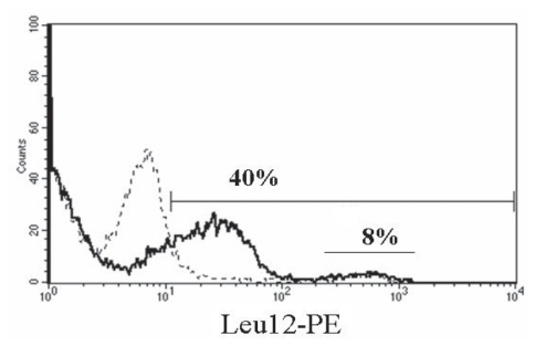
Figure 4. Neuraminidase treatment reveals Leu12 epitopes on MM B cells.
MM PBMC were treated with neuraminidase (solid line) or left untreated (dashed line) as described in methods, followed by staining with Leu12-PE. The marker bar indicates staining above negative control values. The numerical value above the marker bar reports the number of Leu12+ cells after neuraminidase treatment. The Leu12bright peak between 102 and 103 represents expression that is independent of neuraminidase treatment (dotted line), while the lower intensity peak represents Leu12 epitopes revealed by neuraminidase (solid line). PBMC were also stained before and after treatment with fluorescent conjugates of FMC63, B4, CD20 mAbs B1 and rituximab; the staining profiles were comparable before and after treatment for all antibodies except Leu12.

