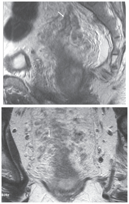
Figure 1.
MR imaging demonstration of extramural venous invasion. T2-weighted imaging in the sagittal and coronal planes demonstrating multiple expanded serpingenous structures (arrows) of intermediate signal intensity emanating from the tumour. This is an example of grade 4 extramural vascular invasion.

