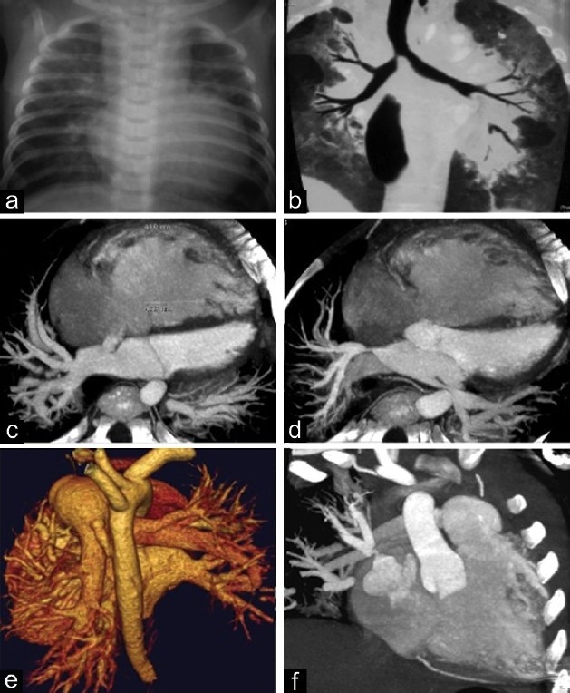
Figure 2.

This figure illustrates the complexity of pulmonary hypertensive vascular disease in a two-year-old infant with bronchopulmonary dysplasia. (a) chest X ray, cardiomegaly and parenchymal lung infiltrates; (b) lung CT scan, showing lung extensive parenchymal damage with areas of atelectasis and emphysema; (c) CT angiogram, showing right ventricular and right atrial dilatation, and atrial septal defect; (d) CT angiogram showing left and right upper pulmonary vein stenosis; (e) reconstructed CT image showing persistent ductus arteriosus; and (f) CT angiogram showing the severe stenosis of right upper pulmonary vein.
