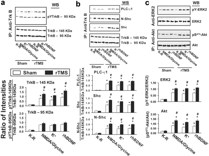
Figure 1.
rTMS treatments increase BDNF–TrkB signaling in prefrontal cortex. a–c, Representative blots (top) with normalized densitometric data (bottom) showing the Western analysis of the effect of 5 d rTMS on tyrosine-phosphorylated (pY) TrkB levels (a); the levels of PLCγ1 and adaptor proteins, shc and N-shc, recruited to TrkB (b); and the levels of activated ERK2 (pY-ERK2) and phosphorylated Akt (pS473-Akt) (c) in response to exogenously added rhBDNF or endogenously released BDNF by K+-depolarization or 10 μm NMDA + 1 μm glycine in the anti-TrkB (a, b) and -ERK2 or Akt (c) immunoprecipitates of prefrontal cortical slice lysates prepared from sham-treated or 5 d rTMS rats. The blots were stripped and reprobed with anti-TrkB (a, b) and -ERK2 or Akt (c) to measure immunoprecipitation efficiency and loading. The densitometric quantification was done on six sham control/rTMS pairs. Data are means ± SEM of ratio of the optical intensity of the pY-TrkB (a) or PLCγ1, Shc, or N-shc (b) to the TrkB band or the pY-ERK2 (c) and pS473-Akt (c) to the ERK2 and Akt, respectively, derived from six independent determinations. *p < 0.01 compared to respective Krebs'–Ringer-treated level in the same group. #p < 0.01 compared to respective response in the sham control group.

