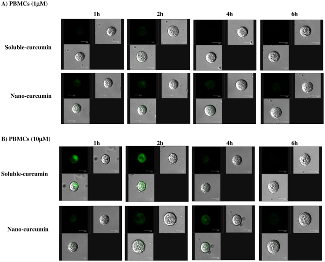
Figure 4. Nanoparticle formulation exhibits increased cellular retention in stimulated PBMCs.
Cells were incubated with 1 µM (Panel A) and 10 µM (Panel B) sol-curcumin and nano-curcumin and examined by confocal microscopy at time points of 1, 2, 4 and 6 h. Each panel contains three images: fluorescence, bright field and merged.

