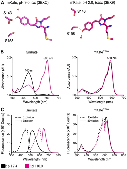
Figure 1. Spectroscopic properties of GmKate and mKateS158A.
A. Position of S158 and S143 in the structures of mKate (PDB codes 3BXC and 3BX9) [5]. Only the chromophore and residues targeted for mutagenesis are shown for clarity. B. Absorbance spectra of GmKate and mKateS158A at pH 7.4 and pH 10.0. The plots were scaled at the same protein concentration. Measurements were carried out at 25°C in buffer containing 150 mM NaCl, 25 mM HEPES or glycine. C. Excitation and emission spectra of GmKate and mKateS158A at pH 7.4 and pH 10.0. Emission spectra for GmKate were recorded at an excitation wavelength of 445 nm (pH 7.4) and 598 nm (pH 10.0), respectively. Emission spectra for mKateS158A were recorded at an excitation wavelength of 588 nm at both pHs.

