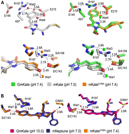
Figure 4. Structural comparison of mKateS158A, GmKate, mKate (PDB: 3BXB) and mNeptune (PDB: 3IP2).
A. Superposition of mKateS158A and GmKate with wild-type mKate [5] at neutral pH. B. Structural comparison of mKateS158A and GmKate (pH = 10.0) with mNeptune [7]. Selected residues in vicinity of the chromophore are shown.

