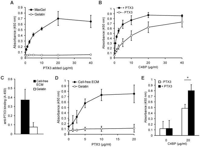
Figure 6. PTX3 recruits C4BP to human fibroblast- and to endothelial cell-derived ECM.
(A) Dose-dependent binding of recombinant human PTX3 to microplate wells coated with human fibroblast-derived ECM (MaxGel™). Gelatin-coated wells served as control. Data shown are means ± SD from three experiments. (B) Dose-dependent binding of purified C4BP to MaxGel™ without or with preincubation with 20 µg/ml PTX3 was measured by ELISA. Data are mean values ± SD of three experiments. (C) PTX3 is present in HUVEC-derived ECM. HUVEC were cultured for 7 days in gelatin-coated 96-well plates, then the cells were detached, and PTX3 which was produced and released by HUVEC and bound to the ECM was detected using an anti-PTX3 mAb. Data shown represent mean + SD of three experiments. (D) HUVEC were cultured for 7 days in gelatin-coated 96-well plates, then the cells were detached and binding of PTX3 to the cell-free ECM was analyzed by ELISA. Gelatin-coated wells incubated with cell-culture medium were used as negative controls. Recombinant PTX3 was added in increasing concentrations and PTX3 binding was detected using a polyclonal anti-PTX3 antibody. Data shown represent mean ± SD of three experiments. (E) Binding of 20 µg/ml C4BP to HUVEC-derived ECM without (white bars) or with preincubation with 20 µg/ml PTX3 (black bars). PTX3 bound to the ECM significantly increased C4BP binding. Data shown are mean + SD derived from three independent experiments. * p<0.05, Student's t-test.

