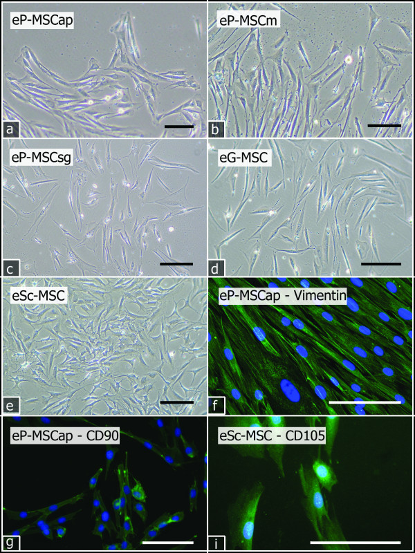
Figure 1.
Primary cells. Reverse phase contrast (a-e) and fluorescent (f) images of cultured primary cells (P0). All primary cells showed fibroblastic morphology and adherence to plastic culture dishes. eP-MSCap (a): day 11; eP-MSCm (b), eP-MSCsg (c), eG-MSC (d): day 18; eSc-MSC (e), day 6. All cells were positive for vimentin (green), here exemplarily shown for eP-MSCap (f). Colony forming cells from passages 2 and 3 stained positive for CD90 (green), shown for eP-MSCap (g) and for CD105 (green), shown for eSc-MSC. Cell nuclei were stained with DAPI (blue). Scale bar = 100 μm.

