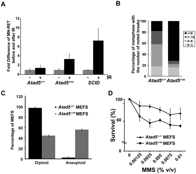
Figure 2. The heterozygous Atad5+/m mutation causes genomic instability in vivo and in MEFs.
(A) Atad5+/m mice exhibited an increase in IR-induced MN-RETs. Wild type (Atad5+/+) and SCID mice were treated with IR and their peripheral blood cells were analyzed in the same manner for comparison. CD71 and propidium iodide (PI) were used to detect RET cells and micronuclei, respectively. Fold induction after IR irradiation in each mouse with a different genotype normalized to MN-RET from unirradiated mice is presented in the graph. Five wild type, five Atad5+/m, and two SCID mice were analyzed. (B) MEFs derived from the Atad5+/m mice produced more chromatid breaks than wild type MEFs in response to MMS treatment. Percentages of metaphases having noted number of breaks are presented as graphs. Noticeable chromatid breaks per cell were quantified for 50 metaphases from both wild type and the Atad5+/m MEFs. (C) MEFs derived from the Atad5+/m mice have a high level of aneuploidy. (D) Survival of wild type and the Atad5+/m MEFs following MMS treatment is displayed as the percentage of viable cells relative to untreated cells. P-values were calculated or each data point as follows, 0.00125% MMS (p = 0.05); 0.0025% MMS (p = 0.002), 0.005% MMS (p = 0.005), 0.0075% MMS (p = 0.09); and 0.01% MMS (p = 0.03), respectively.

