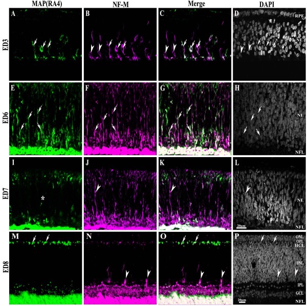
Figure 1.
MAP(RA4) and NF-M expression in the central retina throughout development. Transverse sections of central chick retina at different stages of development processed for double-label immunohistochemistry for MAP(RA4) (green) and NF-M (magenta). Retinal sections are shown with the pigment epithelium at the top and the vitreal surface of the retina at the bottom. (A-D) At ED3 both MAP(RA4) and NF-M are coexpressed in cells located in the most central region of the retina (arrows); a few cells expressing only NF-M were seen at the leading edge of the neurogenic front (arrowheads). (E-H) At ED6 most if not all MAP(RA4) cells are also positive for NF-M. (I-L) By ED7 MAP(RA4) expression is mostly absent from cells in the middle portion of the retina (I; asterisk) while NF-M remains present (J). (M-P) At ED8 expression of both becomes restricted to the nerve fiber layer, GCL, IPL and HCL, with NF-M being also expressed in the bullwhip cells (arrowheads). Arrows indicate cells that are positive for both markers and arrowheads represent cells that are NF-M positive only. Scale bar in L applies to A-L and scale bar in P to M-P. ED: embryonic day; GCL: ganglion cell layer; HCL: horizontal cell layer; INL: inner nuclear layer; IPL: inner plexiform layer; NE: neuroepithelium; NFL: nerve fiber layer; ONL: outer nuclear layer; OPL: outer plexiform layer; RPE: retina pigmented epithelium.

