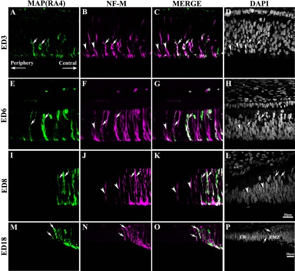
Figure 2.
Progression of MAP(RA4) and NF-M expression towards the periphery during retinal development. Transverse sections of peripheral regions of chick retina at different stages of development processed for double-label immunohistochemistry for MAP(RA4) (green) and NF-M (magenta). Retinal sections are shown with the pigment epithelium at the top and the vitreal surface of the retina at the bottom. As development progresses MAP(RA4) and NF-M expression expands towards more peripheral regions of the retina until they reach the CMZ. At developmental stages between ED3 and ED8 (A-L) NF-M expression was always seen ahead of that of MAP(RA4). By ED18 (M-P) MAP(RA4) is expressed in most if not all cells expressing NF-M in the most peripheral retina. Arrows indicate cells colabeled with MAP(RA4) and NF-M, and arrowheads point at cells that are only positive for NF-M. Asterisks in E-G indicate transverse-sectioned nerve fibers labeled with MAP(RA4) and NF-M. Scale bar in L applies to A-L and scale bar in P applies to M-P. CB: ciliary body; CMZ: ciliary marginal zone; ED: embryonic day; NE: neuroepithelium; RPE: retina pigmented epithelium.

