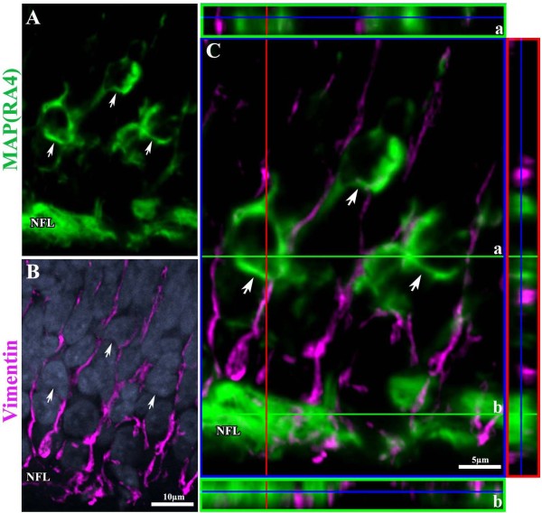
Figure 4.
MAP(RA4) is not expressed in Müller Glial cells. (A-B) Confocal images of a transverse section of an ED6 retina processed by double-label immunohistochemistry for MAP(RA4) (green; A); and (B) vimentin (magenta) and DAPI nuclear staining (blue). (C) Confocal image with orthogonal views depicting no colocalization of MAP(RA4) and vimentin. Arrows indicate MAP(RA4) positive cells. (a) orthogonal view at the level of MAP(RA4) cell bodies; (b) orthogonal view at the level of the nerve fiber layer (NFL).

