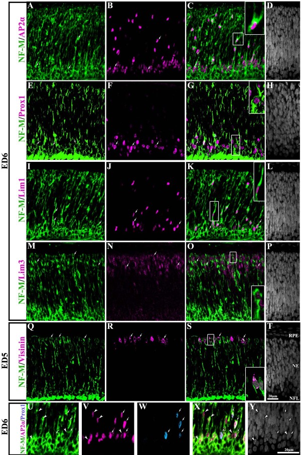
Figure 7.
NF-M is expressed in all neuronal retinal precursor types. Transverse sections of central retina at ED6 (A-P) and ED5 (Q-T) processed for double-label immunohistochemistry for NF-M (green) and AP2α (A-D), Prox1 (E-H), Lim1/2 (I-L), Lim3 (M-P) and visinin (Q-T) all in magenta. NF-M expression was seen in cells positive for markers specific for amacrine cells (A-D), horizontal cells (E-L), bipolar cells (M-P) and photoreceptors (Q-T). Arrows point to cells that are colabeled. Insets represent magnified images of boxed-cells in their corresponding panel. (U-Y) transverse section of an ED6 retina processed by triple-label immunohistochemistry for NF-M protein (green) in combination with AP2α (magenta) and Prox1 (blue). Arrows point to cells that are triple-labeled while arrowheads indicate cells colabeled only with NF-M and AP2α. Scale bar in T applies to panels A-T and scale bar in Y applies to U-Y. NE: neuroepithelium; NFL: nerve fiber layer; RPE: retina pigmented epithelium.

