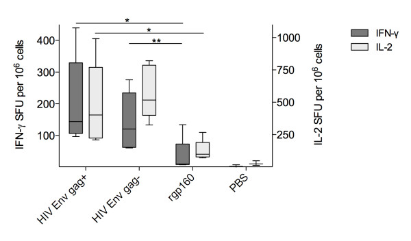
Figure 2.
FLUOROspot of splenocytes isolated from BALB/c mice vaccinated twice with VLPs, microsome-associated env (injecting equal amounts as determined by densitometry) or rgp160 (1 ug/injection) four weeks apart. Box and whiskers plot with 5-95% percentile showing IFN-γ (dark) and IL-2 (light) positive counts in the three. Group medians compared with a Mann-Whitney test * p < 0.05; ** p < 0.01.

