
FIG. 7.
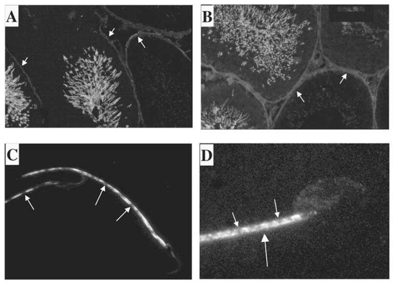
KLC3 localization in testis. The expression pattern of KLC3 protein in seminiferous tubules was examined by immunofluorescence using the mAb raised against KLC3. Frozen rat testicular sections were analyzed using anti-KLC3 mAb (A and B). Tails of KLC3-positive cells can be seen protruding into the lumen. A detailed image of KLC3 in sperm tails was obtained by deconvolution confocal microscopy of rat and mouse epididymal spermatozoa using anti-KLC3 mAb (C and D, respectively). Note that only the midpiece shows KLC3 staining, and that this pattern is not homogenous. Original magnification ×20 (A and B) and ×100 (C and D).