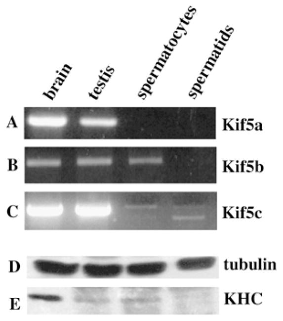
FIG. 8.

Expression pattern of the three known KHC genes in mouse testis. The KHC gene expression in brain, testis, spermatocytes, and spermatids (as indicated) was carried out as described previously by RT-PCR. Primer pairs were specific for Kif5a, Kif5b, and Kif5c (A–C, respectively). Note the absence of Kif5a in male germ cells and of Kif5b in spermatids. Kif5c is expressed at very low levels in spermatids. Western blot analysis of KHC expression in brain, testis, spermatocytes, and spermatids, as indicated. Samples and procedures were standardized for protein content, as indicated by the similar amount of tubulin in the various samples using anti-tubulin antisera (D). The expression of KHC was analyzed using H1 and H2 mAbs, which recognize Kif5b and Kif5c (E). This procedure cannot discriminate between the two proteins, which differ by only 1 kDa. Note the very low level of expression in spermatids.
