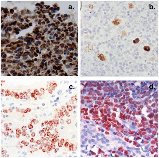
Figure 2. Histological cross sections of typical tumor biopsies used in this study stained for expression of EBV genes.
Note that with the exception of HD less than half of the cells are non-tumor infiltrating lymphocytes. A. Nasopharyngeal carcinoma stained for the EBV nuclear antigen EBNA1. B. Hodgkin's disease stained for EBNA1 Note the sparse appearance of the tumor cells compared to the other tumor types. C. Gastric carcinoma stained for EBNA1 D. Burkitt's lymphoma stained for the EBV encoded small RNAs EBER.

