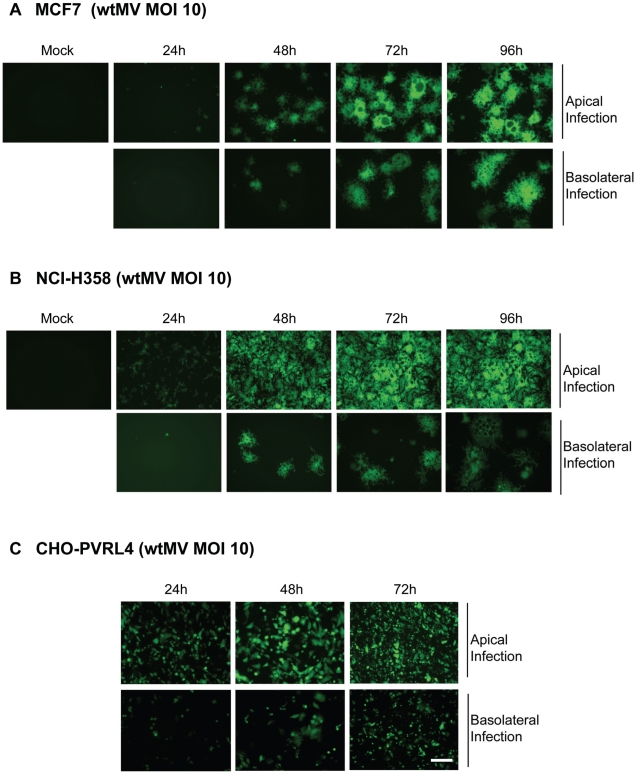
Figure 5. MV infects polarized adenocarinoma cells via either the apical or basolateral surfaces.
Wild type IC323 MV infects (A) MCF7 (breast), (B) NCI-H358 (lung) adenocarcinoma and (C) CHO-PVRL4 cell lines via the apical and basolateral surface in Transwell filter assays. Cells were cultivated in Transwell permeable filter supports at a density of 7.0×10⁵ cells per Transwell filter (24 mm diameter) for 4 days (MCF7 & NCI-H358) or 2 days (CHO-PVRL4). Cells were then infected from either the apical or basolateral side with IC323-EGFP wtMV. At various times post infection fluorescent images were captured. Scale bar = 500 µm.

