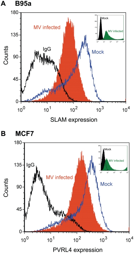
Figure 11. Surface PVRL4 expression is down regulated following wtMV infection.
(A) activated marmoset B-cell line B95a or (B) MCF7 cells were infected with IC323-EGFP wtMV. The fusion inhibitory peptide (FIP) was added after the initial virus infection to prevent syncytia formation. At 48 h post-infection SLAM and PVRL4 surface expression was analyzed by FACS. Blue lines, mock-infected cells stained with alexa anti-SLAM antibody (A) or anti-PVRL4 antibody (B); black lines, mock infected cells stained with the anti-mouse IgG2B isotype control antibody; filled orange histogram, cells infected with IC323-EGFP wtMV (MOI = 10) and stained with anti-SLAM (A) or anti-PVRL4 (B) antibodies, respectively. Alexa fluor conjugated 647 secondary antibodies were used to detect SLAM and PVRL4 surface expression. Insets, level of eGFP positive cells following a 48 h infection with IC323-EGFP wtMV. The filled green histogram represents wtMV-infected cells; black lines represent mock-infected cells.

