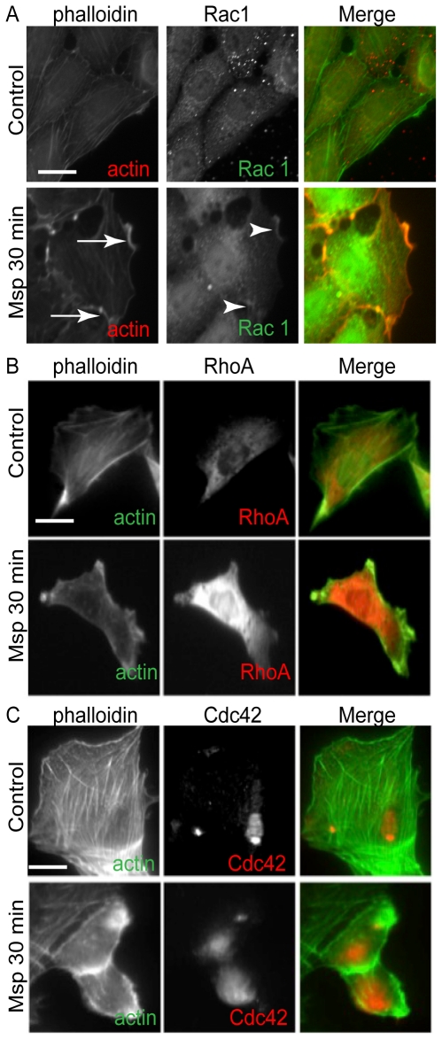
Figure 2. Msp induces re-localization of Rac1 to actin-rich areas.
Immunofluorescence microscopy images of fibroblasts following exposure to Msp. Rac1 (A, arrows) is localized to actin-rich areas (A, arrowheads) at the cell periphery. Msp resulted in changed distribution of both RhoA (B) and Cdc42 (C), however neither localized to areas of remodelled actin. Alexa-phalloidin was used to label F-actin while specific antibodies were used to label each small GTPase. Images are representative of 3 experiments. Bar represents 25 µm.

