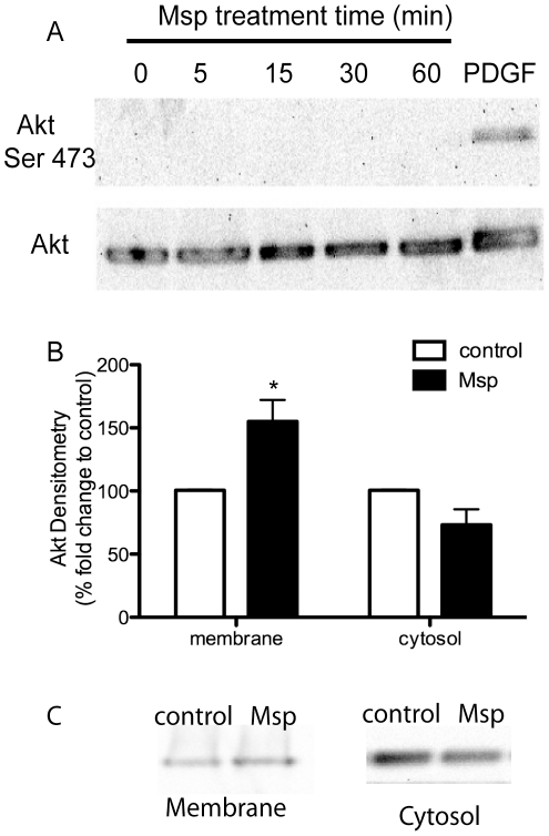
Figure 4. Msp results in Akt translocation but not activation.
(A) Msp treated fibroblasts were analyzed for Akt phosphorylation at serine 473 as a measure of activation. No activation of Akt following Msp exposure was observed by immunoblotting. (B) Densitometry analysis of Akt localization to membrane or cytosol fractions following Msp treatment. Graph represents two immunoblotting experiments (mean ± SEM, * P<0.05). (C) Representative immunoblots demonstrating recruitment of Akt to the membrane following Msp treatment. PDGF treatment of cells serves as a positive control for Akt phosphorylation in panel A.

